
As an avid runner, I like to reward myself with a celebratory meal after a big accomplishment, such as running a marathon. You order a steak and decide to pair it with some shrimp and a baked potato. Every bite is savored as you toast to your accomplishment, noting how it has been perfectly prepared for your liking. Rarely in those moments do we stop and think what testing took place prior to that plate arriving at your table, but behind every high-quality plate of food is rigorous testing working to ensure it’s safe to consume. Nitrofurans, a class of synthetic antimicrobial agents once used in veterinary settings, are now strictly prohibited in livestock and aquaculture due to concerns over their toxic and potentially carcinogenic metabolites. For food testing laboratories, ensuring that your surf and turf is free from these hidden contaminants requires highly sensitive analytical methods capable of detecting nitrofuran metabolites at ultra-trace levels, safeguarding both regulatory compliance and the integrity of every bite.
Nitrofurans history
Nitrofurans are a class of synthetic antimicrobial compounds historically used to treat bacterial infections in livestock and aquaculture. They were first introduced in the 1950s and used in veterinary medicine. Nitrofuran drugs were commonly administered to livestock for several purposes that included:
- Treatment of bacterial infections
- Prevention of disease in intensive production systems
- Growth promotion
- Aquaculture disease control
By the 1960s, the use of these antibiotics spread throughout livestock and aquaculture and became a staple in disease control in the animal population. They were extremely effective against gram-positive and gram-negative bacteria, adding to their growing market value. By the late 1970s, there was increasing concern about the long-term effects of these drugs. Research showed that long after the parent drug was administered to the animal, their metabolites bound to proteins, allowing them to persist in tissues. These metabolites were shown to have mutagenic and carcinogenic properties in animal studies. Concern over their impact on humans quickly spread because these metabolites were still present even after the food had been processed and cooked. As such, by the 1990s, nitrofurans were becoming banned from use in food-producing animals globally. Structurally, these antibiotics feature a 5-nitrofuran ring system consisting of a five-membered furan ring with four carbon atoms and one oxygen atom. A nitro group (-NO2) is attached to each of these nitrofuran rings in the 2-position. Parent compounds are rapidly metabolized by the electron-deficient nitro group, and that instability causes the parent drug to undergo enzymatic reduction through several intermediate steps. With the rapid loss of the parent compound, chromatographic detection will rely on polar metabolites formed (figure 1).
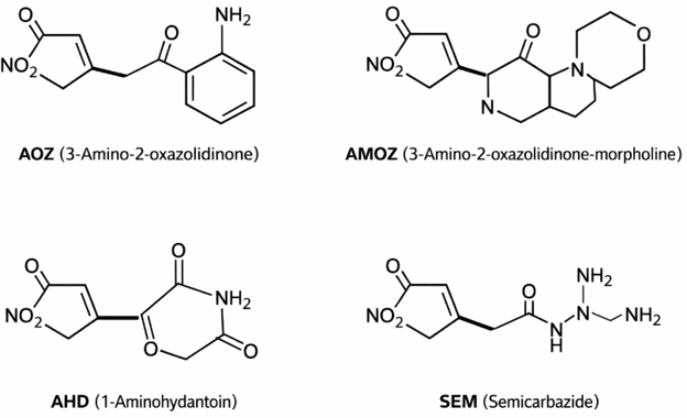
Figure 1: Nitrofuran metabolites
Nitrofuran monitoring applies to a broad range of animal-derived foods such as shrimp, eggs, honey, poultry, and beef. These matrices are protein-rich, requiring efficient hydrolysis and robust cleanup prior to Liquid Chromatography-Tandem Mass Spectrometer (LC-MS/MS) analysis. Nitrofurans are classified as prohibited substances in food-producing animals globally, and regulators established Reference Points for Action (RPA) rather than maximum residue limits. These are highlighted in the table 1 below for each metabolite. Being globally banned means no limit is considered acceptable, so any detection is deemed non-compliant. For AOZ, AMOZ, and AHD, their presence is directly tied to the use of parent compounds. SEM is treated separately due to the known potential for cross-contamination from external sources. This includes environmental contamination in aquaculture systems and contact from different food packaging materials.
Table 1. Regulatory thresholds
|
Analyte |
RPA (µg/kg) |
Regulatory Interpretation |
|
AOZ |
0.5 |
Non-compliant if detected |
|
AMOZ |
0.5 |
Non-compliant if detected |
|
AHD |
0.5 |
Non-compliant if detected |
|
SEM |
0.5 |
Requires context due to background sources |
Monitoring nitrofuran residues remains a critical component of global food safety programs. Because these drugs are banned in food-producing animals, laboratories must detect extremely low concentrations of their metabolites across a wide range of complex matrices. Modern workflows combining acid hydrolysis, derivatization, solid-phase extraction (SPE) cleanup, evaporation, and LC/MS/MS analysis allow laboratories to achieve the sensitivity required for regulatory compliance. There are a variety of methods available for nitrofuran testing, in this article, we will outline the procedure from the Chemical Analytical Manual (CAM) C-011.01 developed by the United States Food & Drug Administration (USFDA)1. This method detects chloramphenicol (a canine antibiotic) as well as nitrofuran metabolites in cobia, croaker, and shrimp. For testing, first, the homogenized samples undergo acid hydrolysis to allow the metabolites to cleave from the animal proteins.
Microwave-assisted derivatization
A 50.0 μL volume of mixed internal standard solution is added to 2.00 grams (± .03) of negative control matrix in a 50 mL centrifuge tube. Approximately 10 mL of 0.125 M HCl is added to each tube. Additionally, 200 μL of 100 mM 2-nitrobenzaldehyde (2-NBA) prepared fresh daily in methanol is added to each vessel. Each vessel is then vortexed (~20 seconds) and placed in the microwave for 6 minutes (5-minute ramp from ambient temperature to 95 ̊C and hold 1 minute). Upon microwave completion, 5 mL of 1.0 M dipotassium hydrogen phosphate is added to each vessel to adjust the pH to 7.3 (± 0.3). If the desired pH is not achieved, the extract can be further adjusted by the addition of diluted sodium hydroxide or hydrochloric acid. Contents are then centrifuged for 10 minutes at 3700g, and the supernatant is subsequently transferred into a 0.45μm PVDF 50 mL filtration tube where it is centrifuged for 5 minutes at 2500g.
Automated positive-pressure SPE cleanup
The 15 mL hydrolyzed sample is prepared for SPE cleanup. These highly polar metabolites must now be isolated from the tissue proteins that they’ve been separated from while being suspended in this highly aqueous solution. A divinylbenzene SPE media is described in the method, making EVOLUTE® EXPRESS ABN an excellent choice. The modified styrene-divinylbenzene polymer sorbent makes it method compliant, and the sorbent is designed to extract acidic, basic, and neutral analytes from a variety of complex matrices. CAM C-011.01 discusses the advantages of using an automated solid-phase extraction (SPE) system as an alternative to liquid-liquid extraction to improve reproducibility and throughput. Depending on the size of the SPE cartridge, the Biotage® Extrahera™ HV-5000 automated SPE system allows for up to 48 samples to be processed at one time. The built-in positive pressure manifold provides highly controlled and consistent nitrogen pressure into each of the sample columns. Coupled with precise pipetting and solvent dispensing, this system delivers exceptional accuracy and reproducibility. The cleanup workflow is conveniently laid out in the method and is easily programmed into the system, as shown in table 2. After the SPE cartridge is conditioned and equilibrated with 3 mL each of ethyl acetate, methanol and water, all 15 mL of the sample is uniformly loaded through the 3 mL cartridge. The aqueous component is transferred to waste, while the compounds of interest are retained to the EVOLUTE® ABN sorbent. 2 mL each of water and a 30% methanol solution are used to wash away any unintended components such as fats, lipids, and proteins. Finally, 3 mL ethyl acetate is used to elute a clean, matrix-free extract containing the analytes of interest. These clean extracts are now ready for evaporation.
Table 2. CAM C-011.01 cleanup
|
Step |
Solvent |
Purpose |
|
Condition |
Ethyl Acetate/methanol/water |
Cleans the sorbent and prepares for sample loading |
|
Load |
Sample extract |
Binds analytes to sorbent |
|
Wash |
Water/30% methanol |
Removes protein matrix residue from the sorbent |
|
Elute |
Ethyl acetate |
Recovers analytes of interest |
Manual positive-pressure SPE cleanup
If a fully automated system isn’t utilized, there are manual manifolds that can still handle the cleanup process, such as the Biotage® PRESSURE+ 48 system. This manual positive pressure system requires more hands-on treatment, so the risk of sample variability will increase, but is still a viable option that is commonly utilized in food testing laboratories.
Sample concentration
A strong organic solvent like ethyl acetate will hinder the polar metabolites from interacting with the LC analytical column, so its evaporation is critical prior to analysis. A nitrogen evaporator is a quick way to conduct this step. The TurboVap® LV evaporator is designed to maximize analyte recovery while minimizing variability. The interchangeable system can easily handle samples in multiple formats, from large volumes of 200 mL all the way down to a few milliliters. This is all achieved from the wide array of manifolds and racks that allow each system to operate in multiple capacities. Samples from the SPE cleanup are collected in 12 x 75 mm borosilicate tubes that are transferred from the Biotage® Extrahera™ HV-5000 to the TurboVap® LV. Extracts are then evaporated to dryness at 55° C and reconstituted with 250 μL of reconstitution solution (40% methanol and 60% 8.5 mM ammonium acetate in 0.1% Formic Acid).
Conclusion
Nitrofuran monitoring remains a key part of maintaining food safety standards globally. Robust workflows must ensure that the polar metabolites are isolated from complex, protein-rich matrices for ultra-trace-level quantification. For this method, microwave derivatization coupled with Biotage® Extrahera™ HV-5000, EVOLUTE® ABN, and TurboVap® LV, produces a reproducible and high-throughput sample preparation solution and meets regulatory requirements for food safety testing.
Learn more about sample preparation strategies for the detection of antibiotics in food matrices here.
Related products
Sources
1. Brian Veach, “Determination of Chloramphenicol and Nitrofuran Metabolites in Cobia, Croaker, and Shrimp Using Microwave-Assisted Derivatization, Automated SPE, and LC-MS/MS–Results from a U.S. Food and Drug Administration Level Three Inter-Laboratory Study,” July 2020, Journal of AOAC International 103(4):1043-1051
